|
|
Pre-natal
Screening During Pregnancy
 A screening test aims to detect a
disease or condition in the early stages
before it causes significant problems, and where treatment can be offered. The
potential benefits of a screening test should outweigh any possible risks from
the test. A screening test aims to detect a
disease or condition in the early stages
before it causes significant problems, and where treatment can be offered. The
potential benefits of a screening test should outweigh any possible risks from
the test.
Both you and your baby will be carefully monitored to ensure your
pregnancy
is progressing well. Part of this prenatal care includes screening tests to help
detect some birth defects. These tests do not usually give a diagnosis, but
indicate whether further, more a accurate tests are necessary. This can be
stressful so you need to be fully informed to keep any development in
perspective. Make sure you know what each test is for, its accuracy, if there
are any risks for you or your baby, and what can be done if the test shows an
abnormality.
This article lists routine
screening tests which should be offered to all pregnant women in and around the
world
Ultrasound scans
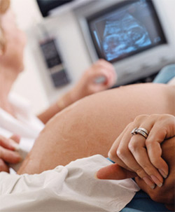
A common screening test during pregnancy is the ultrasound scan. This uses
high-frequency sound waves to display an image of your unborn baby and placenta
on a screen. Ultrasound scans can show your baby's heart beating as early as six
weeks of pregnancy, while later scans can show the details of your baby's face
and genitals -thus revealing its sex-and some developmental abnormalities, such
as spina bifida (where the spinal column does not fuse to protect the spinal
cord, often leading to paralysis and anencephaly (the absence of a skull and
brain).
Ultrasound scanning techniques have come a long way since the first hazy image
produced in the late 1960s. Scans are most useful as a way to answer specific
questions, such as the age of the baby (scans taken in the first trimester can
predict this to within five days), whether he is lying in a
breech or head-down
position, and if his growth rate is normal.
Alpha fetoprotein (AFP) test
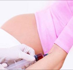
 In recent years obstetricians have developed combinations of blood tests for
pregnant women to try to detect mothers at risk of having a baby with Down
syndrome or spina bifida. Most of these tests include measuring the amount of
alpha fetoprotein (AFP) in the blood. Pregnant women carrying babies with spina
bifida have higher levels of AFP. If a baby has Down syndrome, AFP circulates in
low levels. Mothers-to-be who are shown to have a high risk of a Down syndrome
baby are offered more definitive tests, like
amniocentesis or chorionic villus
sampling. More recently, AFP tests are being replaced by the triple test, which
is taken at 16 weeks of pregnancy. In addition to AFP levels, this test measures
human chorionic gonadotropin (HCG) and estriol level to help identify mothers at
risk of carrying babies with Down syndrome. In recent years obstetricians have developed combinations of blood tests for
pregnant women to try to detect mothers at risk of having a baby with Down
syndrome or spina bifida. Most of these tests include measuring the amount of
alpha fetoprotein (AFP) in the blood. Pregnant women carrying babies with spina
bifida have higher levels of AFP. If a baby has Down syndrome, AFP circulates in
low levels. Mothers-to-be who are shown to have a high risk of a Down syndrome
baby are offered more definitive tests, like
amniocentesis or chorionic villus
sampling. More recently, AFP tests are being replaced by the triple test, which
is taken at 16 weeks of pregnancy. In addition to AFP levels, this test measures
human chorionic gonadotropin (HCG) and estriol level to help identify mothers at
risk of carrying babies with Down syndrome.
The normal levels of these chemicals measured in tests vary considerably with
gestational age, so it's important to remember that, in order to be reliable,
these tests require accurate conception dates. Often, a false alarm is raised
because a woman's pregnancy is inaccurately dated. An early ultrasound scan or
well-kept fertility charts will help you accurately estimate your baby's date of
conception.
Abnormal levels of AFP may signal the following:
-
open neural tube defects (ONTD) such as spina bifida
-
Down syndrome
-
other chromosomal abnormalities
-
defects in the abdominal wall of the fetus
-
twins - more than one fetus is making the protein
-
a miscalculated due date, as the levels vary throughout pregnancy
Amniocentesis
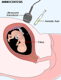
 This involves inserting a long, fine needle through a pregnant woman's abdomen
to draw out some of the amniotic fluid surrounding her unborn baby.
(Amniocentesis is always performed under ultrasound guidance to determine the
position of the baby.) The chromosomal defects can be determined. The sex of
your child can also be detected, which is important if a sex linked inherited
disorder, such as hemophila, is suspected. This involves inserting a long, fine needle through a pregnant woman's abdomen
to draw out some of the amniotic fluid surrounding her unborn baby.
(Amniocentesis is always performed under ultrasound guidance to determine the
position of the baby.) The chromosomal defects can be determined. The sex of
your child can also be detected, which is important if a sex linked inherited
disorder, such as hemophila, is suspected.
Amniocentesis is usually performed at 16 weeks of pregnancy and carries a less
than 1 percent risk of miscarriage-that is, there is more than a 99 percent
chance that you will but the results may take two weeks to come back. This
waiting will usually provide a definite answer to whether their child has a
specific chromosomal disorder, such as Down syndrome.
Chorionic villus sampling (CVS)
 This also tests for genetic and chromosomal abnormalities, but is usually only
undertaken when a specific disorder, such as cystic fibrosis, is suspected. A
fine needle is used to draw out some of the tissue (called chorionic villi)
growing at the edge of the placenta, which contains the same genetic information
as the fetal cells. CVS may be performed vaginally or through the abdomen and is
usually carried out from 10 to 12 weeks of pregnancy. Although it can be carried
out earlier than amniocentesis, there is a very slightly higher risk of
miscarriage or stillbirth. Put more positively, there is a 98 percent chance
that CVS will not cause miscarriage or stillbirth. This also tests for genetic and chromosomal abnormalities, but is usually only
undertaken when a specific disorder, such as cystic fibrosis, is suspected. A
fine needle is used to draw out some of the tissue (called chorionic villi)
growing at the edge of the placenta, which contains the same genetic information
as the fetal cells. CVS may be performed vaginally or through the abdomen and is
usually carried out from 10 to 12 weeks of pregnancy. Although it can be carried
out earlier than amniocentesis, there is a very slightly higher risk of
miscarriage or stillbirth. Put more positively, there is a 98 percent chance
that CVS will not cause miscarriage or stillbirth.
Of course, the bottom line with screening tests is what to do if your baby is
diagnosed with a disorder? With most major abnormalities, medical science can
only offer a termination of the pregnancy. Occasionally new developments in
fetal surgery hit the headlines but, at present, very few birth defects or
inherited problems can be treated in utero. If you could not accept a
termination you may decide that some of these tests are not for you. On the
other hand, just knowing ahead of time may help you prepare for the future. This
is an issue that every couple needs to decide for themselves.
Related Links
|
|
|
|
|


