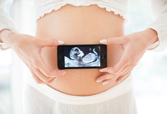
A panel of 15 medical experts from the fields of radiology, obstetrics-gynecology and emergency medicine, convened by the Society of Radiologists in Ultrasound (SRU), has recommended new criteria for use of ultrasonography in determining when a first trimester pregnancy is nonviable (has no chance of progressing and resulting in a live-born baby).
Pelvic ultrasonography and measurement of serum concentration of human chorionic gonadtrophic (hCG) has played a major role in management of early pregnancy problems. In some cases misinterpretation and misuse has been reported. Keeping this in mind new diagnostic guidelines have been published (Oct. 10) in the New England Journal of Medicine, to avoid the possibility of physicians causing inadvertent harm to a potentially normal pregnancy.
The key points made by the expert panel:
- Until recently, a pregnancy was diagnosed as nonviable if ultrasound showed an embryo measuring at least five millimeters without a heartbeat. The new standards raisethe size of embryo to seven millimeters
- The standard for nonviability based on the size of a gestational sac without an embryo has be raised from 16 to 25 millimeters.
- Absence of embryo ≥ 6 weeks after last menstrual period
- Empty amnion (amnion seen adjacent to yolk sac, with no visible embryo)
- Enlarged yolk sac (> 7 mm)
- Small gestational sac in relation to the size of the embryo (< 5 mm difference between mean sac diameter and crown-rump length)
- The commonly used “discriminatory level” of the pregnancy blood test is not reliable for excluding a viable pregnancy.
 The panel has also cautioned physicians against taking any action that could damage an intrauterine pregnancy based on a single blood test, if the ultrasound findings are inconclusive and the woman is in stable condition.
According to Kurt T. Barnhart, MD, MSCE, an obstetrician-gynecologist at the Perelman School of Medicine at the University of Pennsylvania and a member of the SRU Multispecialty Panel “These guidelines represent a consensus that will balance the use of ultrasound and the time needed to ensure that an early pregnancy is not falsely diagnosed as nonviable. There should be no rush to diagnose a miscarriage; more time and more information will improve accuracy and hopefully eliminate misdiagnosis.”
|
Findings diagnostic of pregnancy failure:
Crown-rump length of ≥ 7 mm and no heartbeatMean sac diameter of ≥ 25 mm and no embryoAbsence of embryo with heartbeat ≥ 2 weeks after a scan that showed a gestational sac without a yolk sacAbsence of embryo with heartbeat ≥ 11 days after a scan that showed a gestational sac with a yolk sac |
References
Related Links
- Amniocentesis: learning more about the fetus
- Irregular Menstrual Period
- Top 10 to Minimize the Risk of Miscarriage
Disclaimer
The Content is not intended to be a substitute for professional medical advice, diagnosis, or treatment. Always seek the advice of your physician or other qualified health provider with any questions you may have regarding a medical condition.
