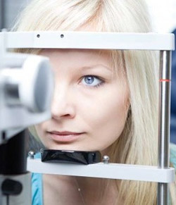
Glaucoma is common in women as they age. Since there are no symptoms in the early stages, having a thorough eye examination is crucial in detecting glaucoma, so you should get yourself or your loved ones checked, especially as you get older.

Glaucoma is detected through an eye exam in which an eye care professional performs some or all of the following:
- Measure your eye pressure (Tonometry): Eye pressure is measured in millimeters of mercury (mm Hg). Normal eye pressure ranges from 12-22 mm Hg, and eye pressure of greater than 22 mm Hg is considered higher than normal. High eye pressure alone does not cause glaucoma. However, it is a significant risk factor. Individuals diagnosed with high eye pressure should have regular comprehensive eye examinations by an eye care professional to check for signs of the onset of glaucoma.
- Inspect the drainage angle of your eye (Gonioscopy): The drainage angle is the point in the eye where the colored part of the eye (iris) and the white covering over the eye (sclera) meet. This is where fluid within the inner eye (which is different from tears that lubricate the eye’s outer surface) drains. Blockage of this angle can lead to increased pressure in the eye, called glaucoma.
- Evaluate your optic nerve (Ophthalmoscopy): This diagnostic procedure helps the doctor examine your optic nerve for glaucoma damage. Eye drops are used to dilate the pupil so that the doctor can see through your eye to examine the shape and color of the optic nerve. The doctor will then use a small device with a light on the end to light and magnify the optic nerve. If your intraocular pressure is not within the normal range or if the optic nerve looks unusual, your doctor may ask you to have one or two more glaucoma exams: perimetry and gonioscopy.
- Dilatation of pupil: Drops are placed in the eyes to dilate, or widen, the pupils. Your eye care professional uses a special magnifying lens to examine your retina to look for signs of damage and other eye problems. A dilated eye exam allows your doctor to check for damage to the optic nerve that occurs when a person has glaucoma. After the examination, your close-up vision may remain blurred for several hours.

It is important to have your eyes examined regularly. Your eyes should be tested:
Anyone with high risk factors should be tested every year or two after age 35. |
You can protect your eye health through regular eye exams to check your optic nerve and eye pressure. If you have already been diagnosed with glaucoma, you can slow the progression of the disease and preserve the vision you still have by following your eye care professional’s instructions for taking your medicine and having regular checkups. If you have already lost some vision, low vision devices may help. Examples include magnifiers, colored lenses, and computer text enlargers.