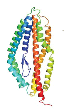
A powerful arm of the immune system is production of antibodies that circulate through the blood and neutralize invading pathogens. Although B cells actually manufacture antibody proteins, the process is aided by neighboring T cells, which shower B cells with cytokines to make them churn out high-quality antibody proteins–and remember how to do so. Given the essential function of “helper” T cells, researchers have long sought to define biological signals that encourage their development. Until now, the best candidates had only minor effects on human immune cells.

Now, a paper published by La Jolla Institute (LJI) researchers in the July 2016 issue of Nature Immunology identifies a human factor that drives maturation of helper T cells known as T follicular helper (Tfh) cells. That factor, a cytokine called activin A, specifically drives maturation of human Tfhs and differs from factors that act similarly in mouse. That work, led LJI vaccine biologist Shane Crotty, Ph.D., fills a knowledge gap by revealing signals that could be therapeutically targeted to either boost immune responses to fight infection or dampen them, in the case of autoimmune disease.
“Most human vaccines work by eliciting an antibody response. For those responses to be of high quality you need Tfh cells,” says Crotty, a professor in LJI’s Division of Vaccine Discovery. “Our study identified an unexpected signaling protein that is very good at inducing maturation of human Tfh cells. Knowing this could help us learn how to design more potent vaccines.”
One reason reaching the goal was challenging is that previously, many research labs, including Crotty’s, had conducted experiments in mouse models and found that factors essential for Tfh differentiation in rodents differed from those regulating human immune cells. So, focusing on human factors only, the Crotty lab set up an unbiased search of 2,000 candidate proteins, an automated strategy called a “high throughput screen.”
Specifically, their task was to comb through a collection or “library” of candidate signaling molecules and test them one by one for whether each could stimulate maturation of cultured immature human Tfh cells. The top candidate emerging from the screen was a secreted protein called activin A, a member of the cytokine family.
Follow-up experiments confirmed that exposure to activin A stimulated immature Tfh cells to express genes encoding receptors and other factors typically seen in mature cells, while fluorescence microscopy revealed activin A protein in lymphoid tonsil tissues near sites where Tfh differentiation took place and in close proximity with B cells. That circumstantial evidence proved that activin A was produced in the “right place.”
Further comparisons explored and confirmed the rodent/human divergence that had proven to be such an obstacle. Incubation of immature mouse T cells with activin A did not in fact prompt their maturation, while treating immature Tfhs from macaque monkeys with activin A did.
The discovery of a uniquely primate mechanism is intriguing from an evolutionary point of view, as biochemical pathways regulating immune activities are often highly conserved across mammals. “Mice are extremely useful in biomedical research in helping us understand how genes and proteins work,” says Crotty. “But occasionally, one sees species differences, and Tfh cell differentiation was one of those cases.”
Finally, the team showed that for activin A to stimulate Tfh maturation it must activate downstream DNA-binding molecules called SMAD2/3. They discovered this by treating immature Tfh cells with the drug Galunisertib, which muffles SMAD2 signaling, and found that cells became refractory to activin A signals. This experiment not only defines a biochemical pathway but means that a drug exists to potentially block T cell “help” by keeping Tfh cells in an immature state–the most obvious application being to autoimmune disease.
“Many autoimmune diseases exhibit excessive generation of Tfh cells, which can lead to formation of auto-reactive antibodies,” says Michela Locci, Ph.D., the study’s lead author and a postdoctoral fellow in the Crotty lab. “In at least two of them–rheumatoid arthritis and systemic lupus erythematous–the level of activin A in blood is higher than normal, suggesting that it contributes to dysregulated Tfh differentiation in these diseases.” Locci finds these associations promising but cautions that it is too early to say whether Galunisertib, which is being tested as an anti-cancer drug in clinical trials, could normalize excess Tfh activity in autoimmune disease.
Overall, the Crotty lab’s predominant interest is in antibody-based immunity as it relates to vaccine development. One way that vaccines work is by establishing long-lived memory B cells that when exposed to a deadly pathogen recall that they have seen the invader before (albeit in a harmless vaccine form) and are prepared to make antibodies. When Tfh cells were discovered in 2001, researchers soon realized that they were necessary for both establishing those memory B cells and instructing them in how to make high affinity antibodies.
“Generating more Tfh cells could promote more potent B cell responses and increase production of long-lived memory cells,” says Crotty. “From a vaccine design perspective it might be desirable to encourage Tfh cell development in vivo since generation of a protective vaccine requires antibody production.” Knowing that activin A is the chief source of “encouragement” could lead his lab to a new search, this time for small molecules potentially useful to turn activin A on at the time of vaccination, to instill more long-lasting memories in B cells.