For people who have gone blind from degenerative eye diseases like macular degeneration and retinitis pigmentosa. Both diseases damage the eyes’ photoreceptors, the cells at the back of the retina that perceive light patterns and pass them on to the brain in the form of nerve impulses, where the impulse patterns are then interpreted as images.

The Argus II system takes the place of these photoreceptors.
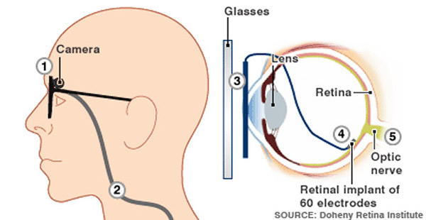
Second Sight’s retinal prosthesis consists of five main parts:
- A digital camera that’s built into a pair of glasses. It captures images in real time and sends images to a microchip.
- A video-processing microchip that’s built into a handheld unit. It processes images into electrical pulses representing patterns of light and dark and sends the pulses to a radio transmitter in the glasses.
- A radio transmitter that wirelessly transmits pulses to a receiver implanted above the ear or under the eye
- A radio receiver that sends pulses to the retinal implant by a hair-thin implanted wire
- A retinal implant with an array of 60 electrodes on a chip measuring 1 mm by 1 mm
The Bionic Eye

The entire system runs on a battery pack that’s housed with the video processing unit.
- When the camera captures an image — of, say, a tree — the image is in the form of light and dark pixels.
- It sends this image to the video processor, which converts the tree-shaped pattern of pixels into a series of electrical pulses that represent “light” and “dark.”
- The processor sends these pulses to a radio transmitter on the glasses, which then transmits the pulses in radio form to a receiver implanted underneath the subject’s skin.
- The receiver is directly connected via a wire to the electrode array implanted at the back of the eye, and it sends the pulses down the wire.
- When the pulses reach the retinal implant, they excite the electrode array. The array acts as the artificial equivalent of the retina’s photoreceptors. The electrodes are stimulated in accordance with the encoded pattern of light and dark that represents the tree. The electrical signals generated by the stimulated electrodes then travel as neural signals to the visual center of the brain by way of the normal pathways used by healthy eyes — the optic nerves. In macular degeneration and retinitis pigmentosa, the optical neural pathways aren’t damaged. The brain, in turn, interprets these signals as a tree and tells the subject, “You’re seeing a tree.”
It takes some training for a person who has undergone this operation to actually see a tree. At first, they see mostly light and dark spots. But after a while, they learn to interpret what the brain is showing them, and they eventually perceive that pattern of light and dark as a tree.
| On March 24, 2015 a cutting-edge procedure was carried out by Dr. Gregg T. Kokame of the Hawaii Eye Surgery Center. This surgery comes two years later after the first bionic implant of a prototype conducted by researchers from Bionic Vision Australia. After the four-hour surgery a 72-year-old woman who had been blind for two years said that she had started to see shades of grey. |
Ref:
Related Links
Disclaimer
The Content is not intended to be a substitute for professional medical advice, diagnosis, or treatment. Always seek the advice of your physician or other qualified health provider with any questions you may have regarding a medical condition.

